

Per a prescription order, a formulation can be compounded to contain the proper combination of active ingredients, in the most appropriate base, to treat a specific type of wound. We customize medications to meet each individual’s specific needs.
For example, the choice of cream, ointment, or gel can be clinically significant. Each time a wound needs to be cleaned, there is the potential for disruption of new tissue growth. Gels, which are more water soluble than creams or ointments, may be preferable for wound use because a gel can be rinsed from the wound by irrigation. Ointments may contain polyethylene glycol (PEG), which can be absorbed from open wounds and damaged skin. If the wound is quite large and too much PEG is absorbed, it can lead to renal toxicity.
Another useful dosage form is the “polyox bandage” – which can be puffed onto a wound and will adhere even if exudate is present. A polyox bandage can be compounded to contain the active ingredient(s) of your choice.
Overview
Phenytoin has been used topically to speed the healing of decubitus ulcers, pressure sores, venous stasis and diabetic ulcers, traumatic wounds, skin autograft donor sites, and burns. Ketoprofen may be used to control inflammation and pain, lidocaine provides topical anesthesia, and pentoxifylline may improve microcirculation at the wound margins and promote healing of the injured area. Misoprostol, a prostaglandin analog, is often included in wound care formulations to promote healing.
Debridement of necrotic eschar with 40% urea paste may also speed healing. Medications which improve capillary blood flow can be added to a compounded medication to enhance circulation at the wound margins and promote healing of the injured area.
Topical Phenytoin for Wound Healing
The stimulatory effect of orally administered phenytoin on gingival tissue prompted its assessment in wound healing. Phenytoin may promote wound healing by a number of mechanisms, including stimulation of fibroblast proliferation, facilitation of collagen deposition, glucocorticoid antagonism, and antibacterial activity. Phenytoin has been used topically in the healing of pressure sores, venous stasis and diabetic ulcers, traumatic wounds, skin autograft donor sites, and burns.
Rhodes et al compared the healing of stage II decubitus ulcers with topically applied phenytoin and two other standard topical treatment procedures in 47 patients in a long-term care setting. Ulcers were examined for the presence of healthy granulation tissue, reduction in surface dimensions, and time to healing. Topical phenytoin therapy resulted in a shorter time to complete healing and formation of granulation tissue when compared with DuoDerm dressings or triple antibiotic ointment applications. The mean time to healing in the phenytoin group was 35.3 +/- 14.3 days compared with 51.8 +/- 19.6 and 53.8 +/- 8.5 days for the DuoDerm and triple antibiotic ointment groups, respectively. Healthy granulation tissue in the phenytoin group appeared within 2 to 7 days in all subjects, compared to 6 to 21 days in the standard treatment groups. The phenytoin-treated group showed no detectable serum phenytoin concentrations.
Anstead et al. described a patient with a massive grade IV pressure ulcer that was unresponsive to conventional treatment, yet responded rapidly to treatment with topical phenytoin. Song and Cheng reported phenytoin improved wound breaking strength in normal and radiation-impaired wounds. The results of their study indicated that topical phenytoin accelerated normal and irradiation-impaired wound healing by increasing the number of wound macrophages and improving the macrophage function. Pendse et al evaluated the effectiveness of topical phenytoin in healing chronic skin ulcers in a controlled trial of 75 inpatients. At the end of the fourth week, 29 of 40 phenytoin-treated ulcers had healed completely versus 10 of 35 controls. They concluded: “topical phenytoin appears to be an effective, inexpensive, and widely available therapeutic agent in wound healing.”
The effectiveness of topical phenytoin as a wound healing agent was compared with that of OpSite and a conventional topical antibiotic dressing (Soframycin) in a controlled study of 60 patients with partial-thickness skin autograft donor sites on the lower extremities. Mean pain scores were lower and mean time to complete healing (complete epithelialization) was best in the phenytoin-treated group (6.2 +/- 1.6 days). Topical phenytoin compared very favorably with, and in some aspects was superior to, occlusive dressings.
The efficacy of topical phenytoin in the treatment of diabetic foot ulcers was evaluated in a controlled inpatient study. Fifty patients were treated with topical phenytoin, and 50 patients received dry sterile occlusive dressings. Both groups improved, but the ulcers treated with topical phenytoin healed more rapidly. Mean time to complete healing was 21 days with phenytoin and 45 days with control.
No study reported any significant adverse effects secondary to topical phenytoin therapy.
Ann Pharmacother 2001 Jun;35(6):675-81
Biochem Pharmacol 1999 May 15;57(10):1085-94
Ann Pharmacother 1996 Jul-Aug;30(7-8):768-75
Int J Dermatol 1993 Mar;32(3):214-7
Chung Hua I Hsueh Tsa Chih 1997 Jan;77(1):54-7
Burns 1993 Aug;19(4):306-10
Diabetes Care 1991 Oct;14(10):909-11
Benzoyl Peroxide for Treatment of Decubitus Ulcers
Benzoyl peroxide is a powerful oxidizing agent with broad spectrum germicidal activity and good liposolubility. Therefore, it may represent a good agent for prevention of wound infection in areas with high density of sebaceous glands. Topical treatment of pressure sore with 20% benzoyl peroxide in O/W emulsion yielded very satisfactory results. In another study, 10% benzoyl peroxide gel was used prophylactically once a day for 7 days before surgery. The researchers concluded that topical benzoyl peroxide is an efficacious, harmless, and inexpensive agent for prevention of wound infections in seborrheic regions.
Med Cutan Ibero Lat Am 1988;16(5):427-9
J Dermatol Surg Oncol 1994 Aug;20(8):538-40
Arch Dermatol. 2001 Oct;137(10):1288-90
Numerous topical preparations containing cholestyramine or sucralfate (creams, adhesive pastes, enemas, suppositories) have been used for their protectant properties or for treatment of a variety of dermatologic and mucosal problems, including oral and esophageal ulcers, peristomal and perineal excoriation, decubitus ulcers, and radiation-induced rectal and vaginal ulcerations, and second and third degree burns.
Ann Pharmacother 1996 Sep;30(9):954-6
Dis Colon Rectum 1987 Feb;30(2):106-7
Clin Exp Dermatol. 2000 Nov;25(8):584-8
Burns. 2001 Aug;27(5):465-9
Odor from malignant cutaneous wounds, ulcerated tumors, some pressure ulcers, and fungating tumors can cause great distress and embarrassment for patients. Topical metronidazole is one medication that has been used to eliminate this odor, greatly improving the patient’s quality of life. Exudate and associated cellulitis may also decrease significantly with appropriate topical therapy.
Malignant cutaneous wounds are emotionally traumatic and difficult to manage lesions which occur secondary to infiltration of cancer into the skin. They occur in patients with end-stage disease and are highly exudative, malodorous, and bleed easily. Quality of life is the goal for treatment, which includes radiation, chemotherapy, surgery, and local wound care. Odor is addressed with varying levels of success through wound cleansing, external deodorizers, charcoal-impregnated dressings, topical antimicrobial therapy, and metronidazole. Exudate is managed with highly absorbent dressing materials, topical steroids or hyoscine (a drying agent). Light bleeding is controlled with local pressure and hemostatic dressings; heavier bleeding may require ligation or cauterization. Cosmetic appearance and other psychosocial issues must be assessed on an ongoing basis. Creative dressing techniques can help restore the look of symmetry to the patient’s body. Effective wound management, debridement, and antimicrobial theray can reduce the risk of infection. Wound cleansing, through irrigation or flushing, should not cause pain, further trauma or bleeding. Dressings should maintain a moist wound environment and not traumatize the wound upon removal. A protocol is included which can be individualized to the needs of each patient and addresses assessment, interventions, patient teaching, documentation, and expected outcomes.
Improving the appearance of scarred skin is not easy. With possibly the exception of young boys, ages 10 to 15, who proudly display theirs as a badge of honor, scars are largely regarded as cosmetically undesirable. Fireside Pharmacy now offers PracaSil™ Plus to serve not only as a standalone cream that can help lighten scars, but also as a base in which your practitioner can incorporate active pharmaceutical ingredients (APIs).
PracaSil™-Plus is an ideal choice for new scars, old scars, surgical scars, keloids, stretch marks, acne scars and any skin conditions. In order to determine which APIs to ask your doctor about first you must identify the type of scar in question.
Scars generally fall into three categories:
- Hypertrophic: Excessive scar tissue that protrudes above the height of normal skin. Scars do not extend beyond the confines of the original wound.
- Keloid: Excessive scar tissue that protrudes above the height of normal skin. More aggressive compared to hypertrophic; scars continually grow and invade the normal surrounding tissue.
- Flat (atrophic): Less aggressive – usually stretch marks and acne scars.
Hypertrophic Scars

Hypertrophic scarring usually occurs after surgery, serious burns, or trauma that involves moderate to extreme penetration into the dermal tissue. For instance, wounds that experience a moderate to an extreme amount of inflammation are more likely to become hypertrophic. Usually within four weeks of the event you will notice hypertrophic scarring.
Both hypertrophic and keloid scars are the result of excessive collagen produced in the healing process. Due to the excess scar tissue, scars will appear raised, thick, and darker than typical scars.
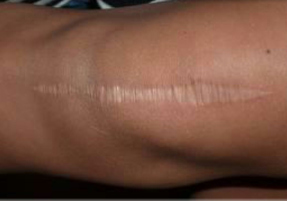
Keloid Scars
keloid scarring: notice the scar tissue spreading
Keloid development is much more problematic. The location of these scars is predominately formed on the earlobes, shoulders, anterior chest, upper arms, or cheeks. The inflammatory response seems to be less of a factor than on where the scar is actually located.
Unlike hypertrophic scars that become prominent as early as four weeks after trauma, keloids often develop months or years after dermal tissue penetration and show very little tendency to regress over time. And while hypertrophic scars remain confined to the area of the original wound, keloids extend into the surrounding tissue. PracaSil-Plus can aid in the prevention of excessive collagen production which causes these disorders.
Actives for Keloid and Hypertrophic Scars
APIs such as captopril, EGCg, tamoxifen, pentoxifylline, imiquimod, and tranilast are usually used to reduce the excessive collagen deposits these scars create. These ingredients have the greatest benefit in the early phase of scar development.
For old keloid and hypertrophic scars, steroids have been selected to reduce inflammation and bulk tension in skin tissue. Collagenase and hyaluronidase can also be incorporated into compounds to reduce scar components.
Flat Scars
Flat scars, stretch marks, and acne scars do not have the same amount of inflammation or collagen deposits as keloid and hypertrophic scars. The main focus on healing flat scars should be to target the gradual renewal of the skin. Tretinoin is a useful agent, as it encourages skin turnover. The desired result is a renewed skin layer that has better character and quality than the existing scar tissue. Additionally, sodium hyaluronate and aloe may be included to increase the moisture content and to soothe the area during this renewal process.
Regardless of the type of scar, the length of therapy generally depends on how long the scar has existed. Older scars will generally require longer therapy. New scars may begin to fade in as little as one or two months, while older scars may take several months to show marked improvement. Call Fireside Pharmacy today and talk to one of our pharmacists about how you can start making your scars fade away today.


